
Every instrument below has a 3D model: press Rotate in 3D, then drag or use the arrow keys to turn it and scroll or pinch to zoom.
Read this first
- The pictures are synthetic. The project built every model from simple shapes in Blender (the script); they are not photographs and not copies of any manufacturer’s product. Real instruments differ in colour, jaw shape and shaft width, so learn the shape first and the colour second.
- Real frames are harder. Smoke, blood, glare, motion blur and the edge of the scope’s field of view all change how an instrument looks. The synthetic frames here show the idea of each rule, not its hardest case.
- The rules are a draft. Class names and colours match the project’s CVAT label set. Defining a class is the Clinical Lead’s decision; until the rules below are approved, the instructions in your CVAT task take precedence over this page.
Drawing an instrument mask
- One shape per instrument. Two graspers in view are two shapes, both labelled
grasper. - The whole visible instrument. Tip or jaws, joint and shaft, up to the edge of the image. The black insulated shaft belongs to the instrument too.
- Only what you can see. Where tissue, blood or another instrument covers part of it, do not draw underneath: the mask may break into pieces, and that is correct.
- Not the surroundings. Tissue held in the jaws, the instrument’s shadow, its reflection on wet tissue, smoke at the tip and the trocar it passes through are not part of the instrument.
- When unsure, say so. If you cannot tell which instrument it is, add the
disputedtag to the frame instead of guessing. It goes to the Clinical Lead, and the guideline improves.
Grasper
CVAT label: grasper
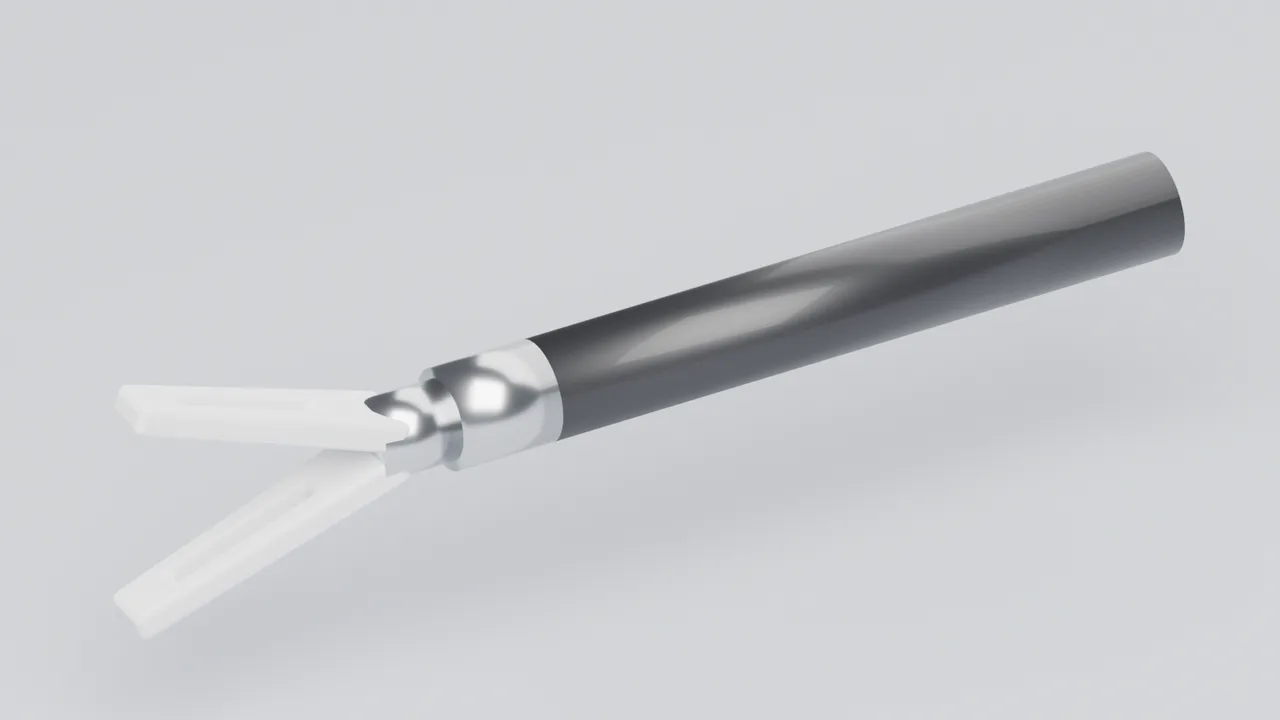
The grasper holds and lifts tissue. In a cholecystectomy it usually holds the gallbladder up and out of the way while another instrument dissects, so it is in view for much of the operation.
- Look for: two straight jaws of the same length that open symmetrically from a joint, often with a window (fenestration) and ridges on the inside; a black insulated shaft about 5 mm across.
- Do not confuse with: the bipolar forceps (slender, often curved jaws with coloured insulation), scissors (thin blades that cross) or the clip applier (a wider shaft and a clip between the jaws).
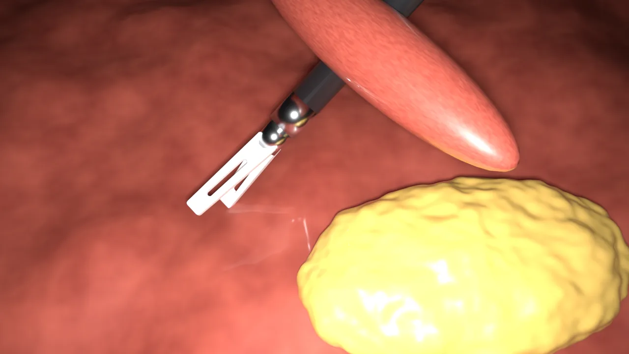
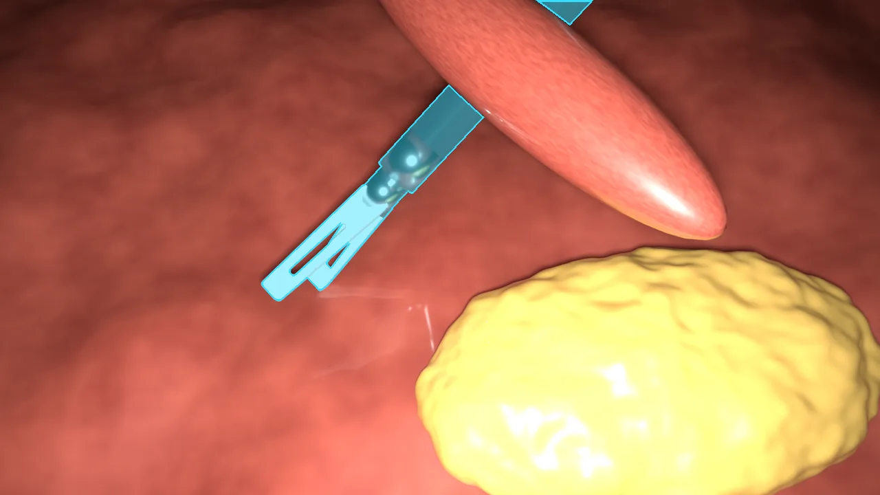
- The mask: jaws, joint and shaft as one shape. Tissue held between the jaws is not part of the grasper.
In the synthetic frame a fold of tissue lies across the shaft. The mask stops on each side of the fold and is not joined underneath it: draw only what you can see.
grasper colour.Bipolar forceps
CVAT label: bipolar
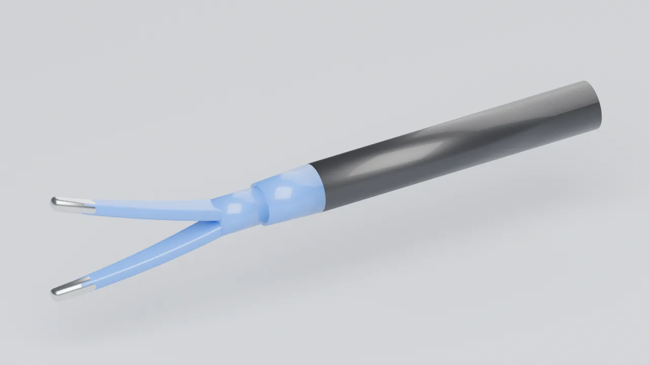
Bipolar forceps pass current between their two jaws to seal small vessels and stop bleeding.
- Look for: slender jaws, often curved, with insulation in a colour (blue on many models) and bare metal at the very tips.
- Do not confuse with: the grasper, whose jaws are broader, straight and usually all metal. Colours differ between manufacturers, so judge by shape first and colour second.
- The mask: both jaws, the insulated joint and the shaft. Smoke or a spark at the tips is not the instrument.
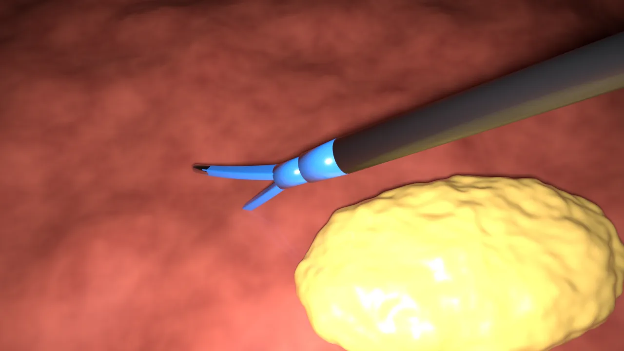
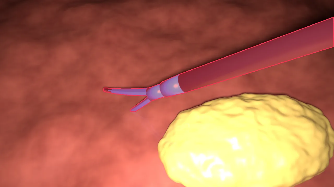
bipolar colour.L-hook
CVAT label: hook
The L-hook is a monopolar electrode used to dissect tissue with current. In CholecSeg8k this class is called L-hook electrocautery.
- Look for: a thin metal wire that leaves a tapered insulated shaft and bends through a right angle at the end.
- Do not confuse with: the irrigator, a blunt tube with no bend. When the bend points straight at the camera the hook can look like a short straight wire, so follow it over neighbouring frames.
- The mask: the shaft, the taper and the whole wire, even where it is only a few pixels wide. The smoke it makes and its glare on wet tissue are not part of it.
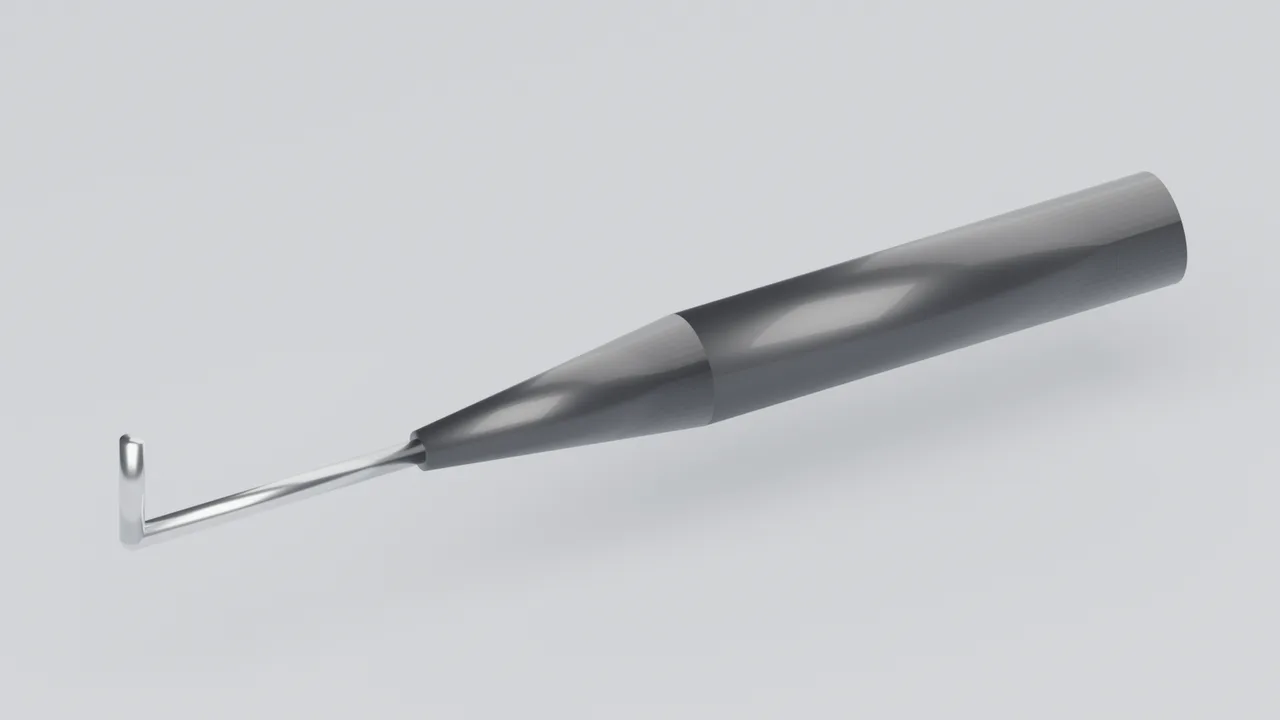
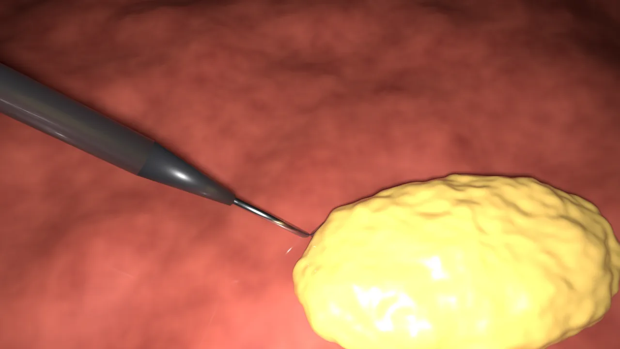
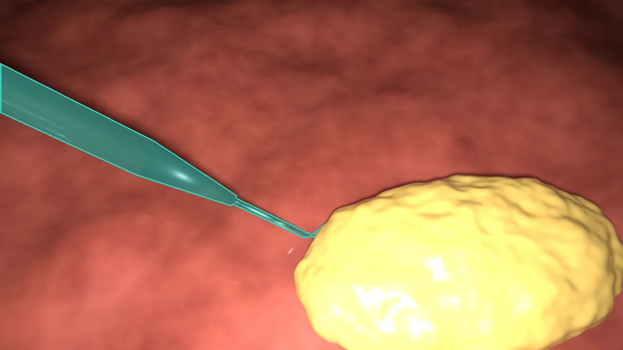
hook colour.Scissors
CVAT label: scissors
Scissors cut: in a cholecystectomy, mostly the cystic duct and the cystic artery between clips.
- Look for: two thin blades, usually curved to one side, that cross each other at a pivot near the tip.
- Do not confuse with: the grasper. Grasper jaws meet face to face; scissor blades slide past each other and have no window.
- The mask: both blades, the pivot and the shaft. The cut tissue is not part of the scissors.
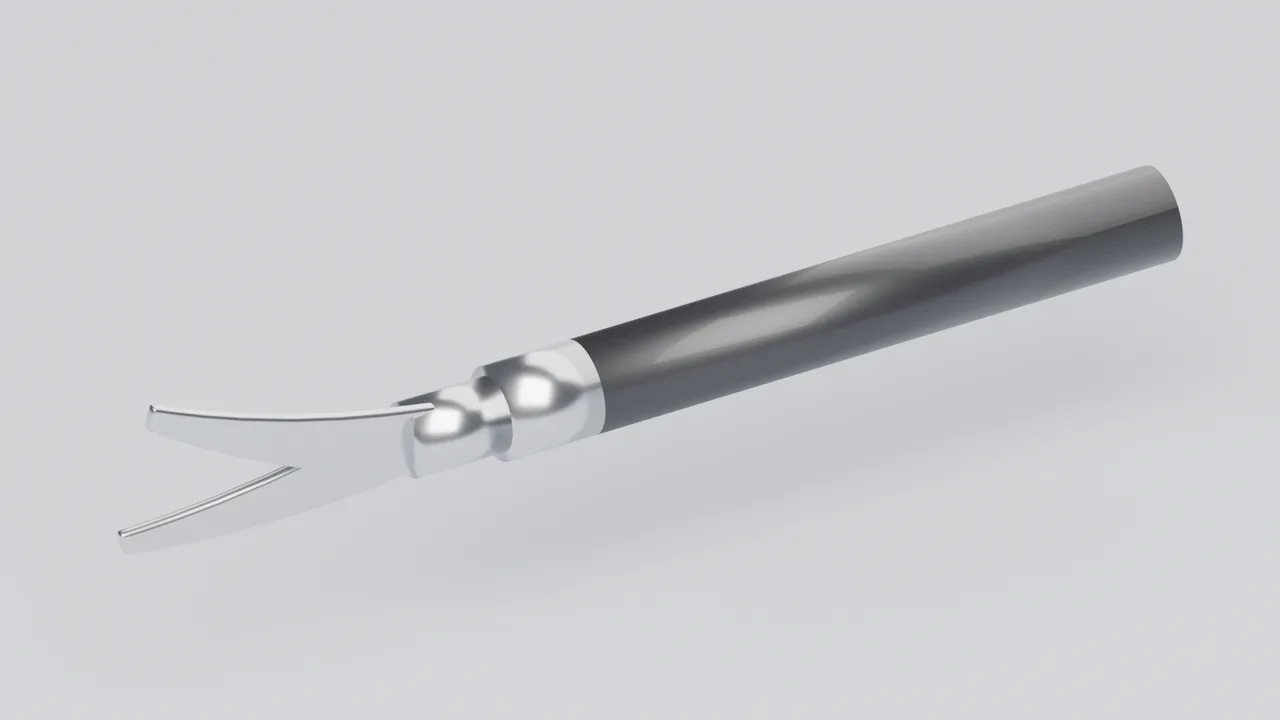
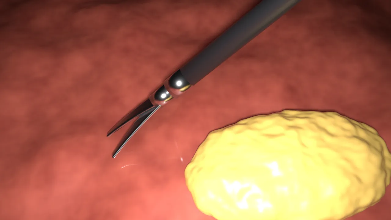
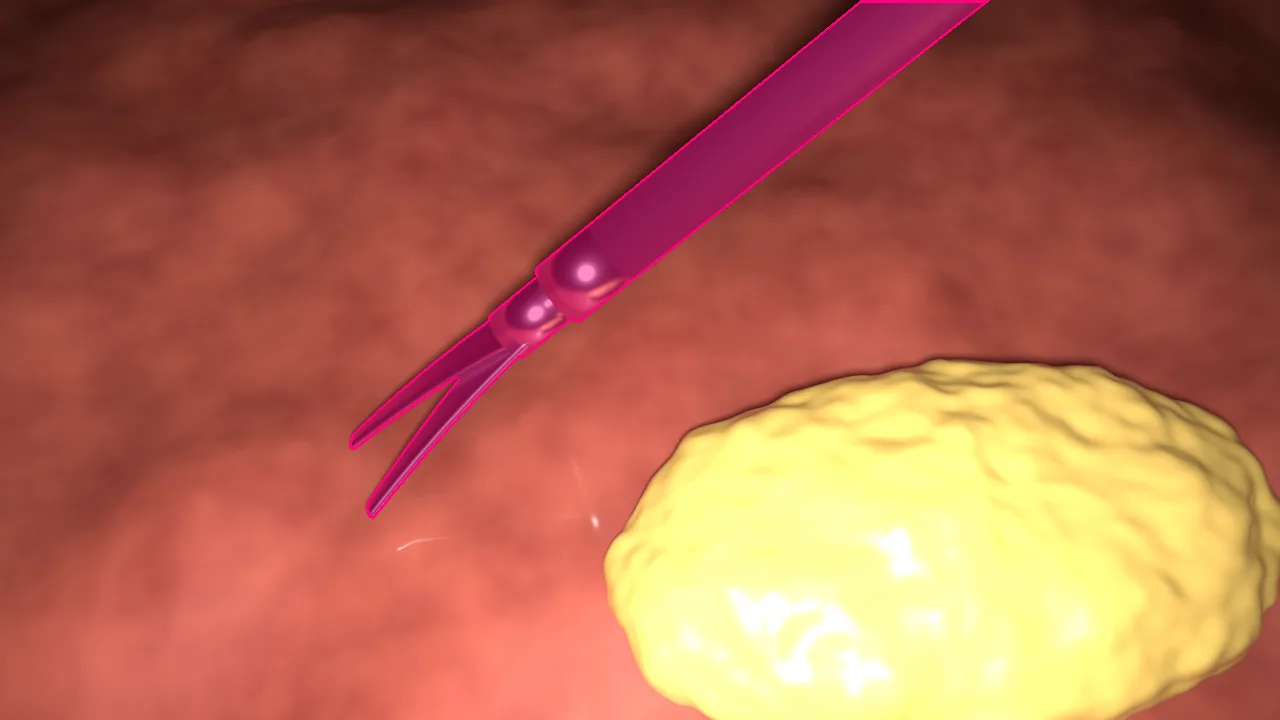
scissors colour.Clip applier
CVAT label: clipper
The clip applier closes the cystic duct and the cystic artery with clips before they are cut.
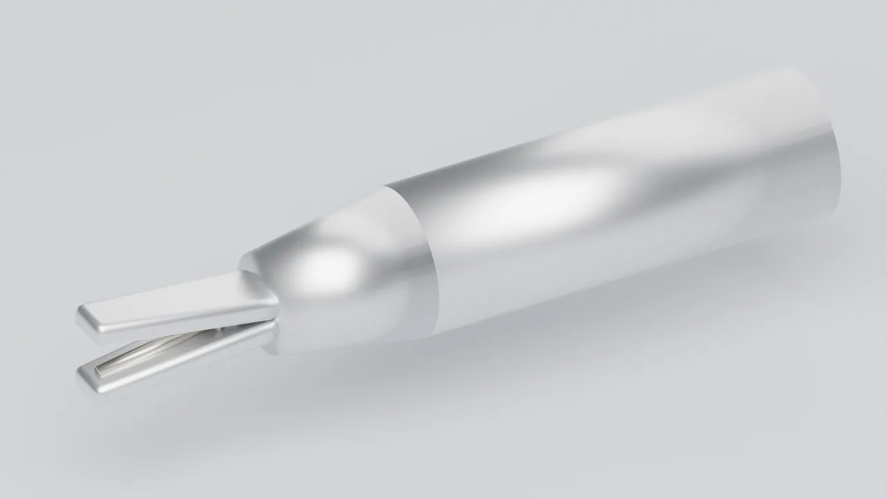
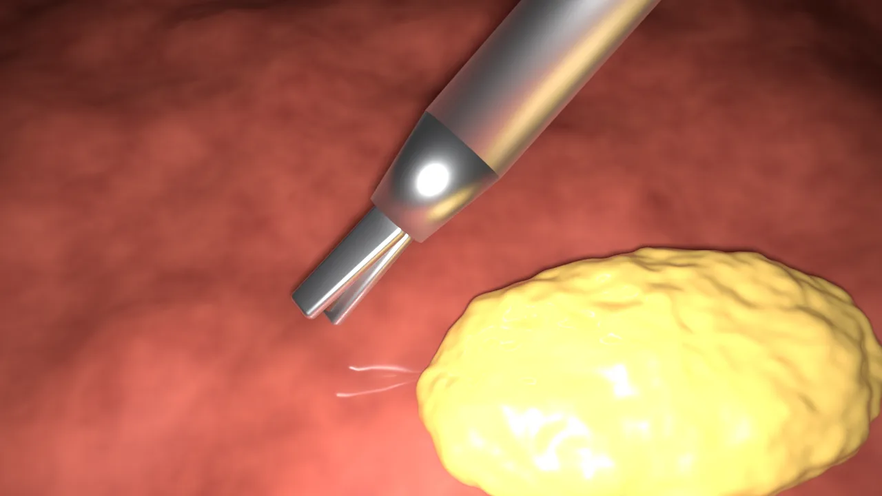
- Look for: a wider shaft than the other instruments (often 10 mm rather than 5 mm), broad jaws that close in parallel, and a clip loaded between them.
- Do not confuse with: the grasper. A clip in the jaws, or a clip left behind on tissue, is the strongest clue.
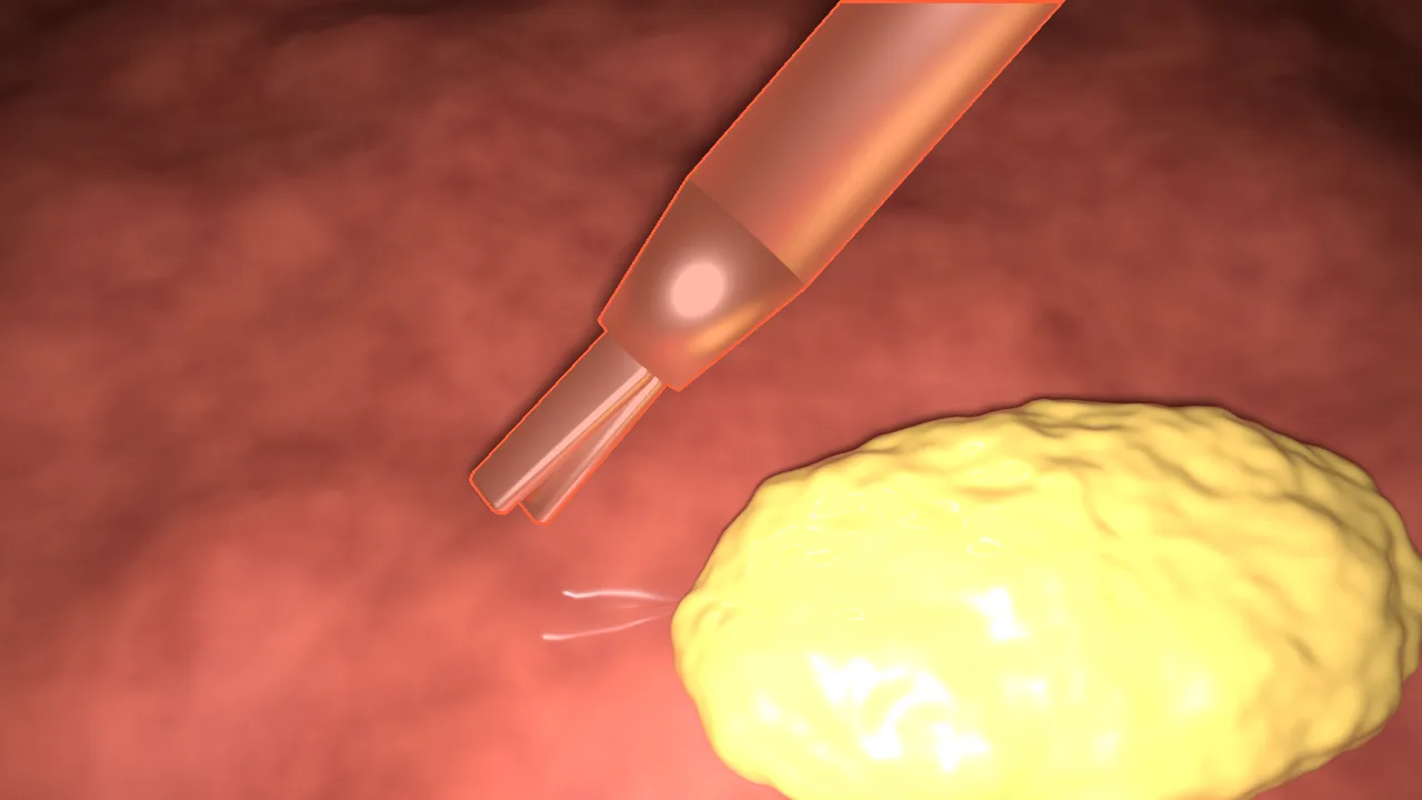
- The mask: jaws, the loaded clip and the shaft. Clips already placed on tissue stay behind when the instrument leaves; they are not
clipper, and the label set has no class for them yet.
clipper colour.Suction-irrigator
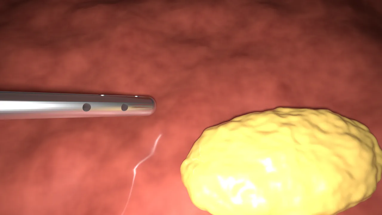
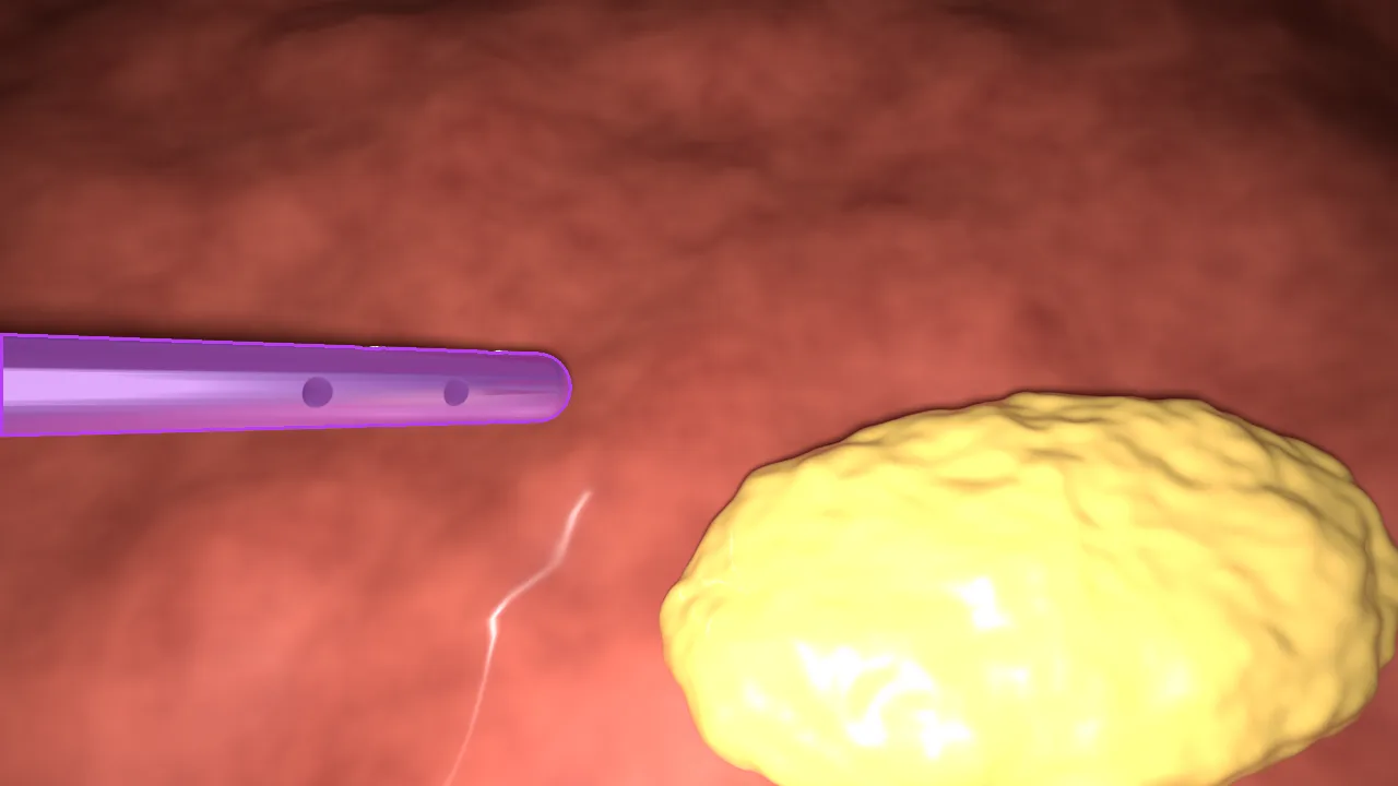
CVAT label: irrigator
The suction-irrigator washes the operating field and sucks out blood, fluid and smoke.
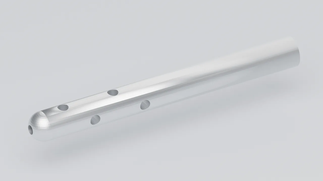
- Look for: a plain metal tube with a blunt rounded end and side holes near the tip; it has no jaws and no bend.
- Do not confuse with: the L-hook (a thin wire with a right-angle bend) or the bare shaft of another instrument whose tip is out of view.
- The mask: the whole visible tube. Fluid coming out of it and blood being sucked in are not part of it.
irrigator colour.Specimen bag
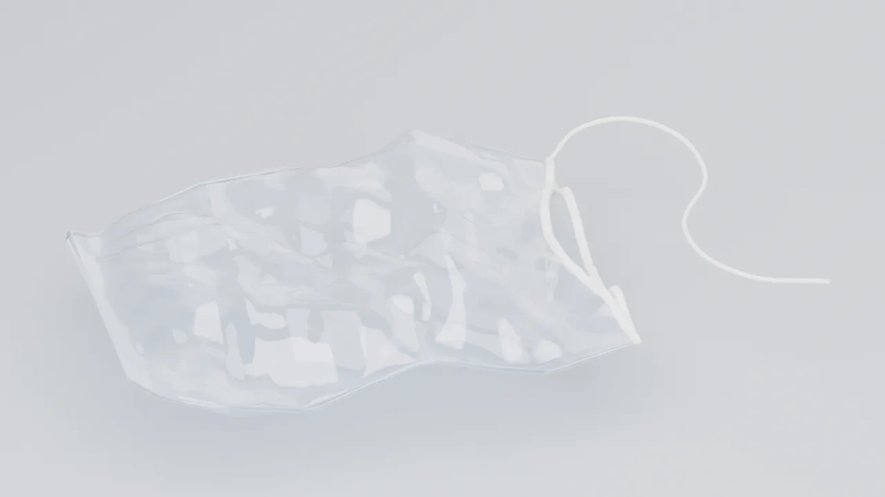
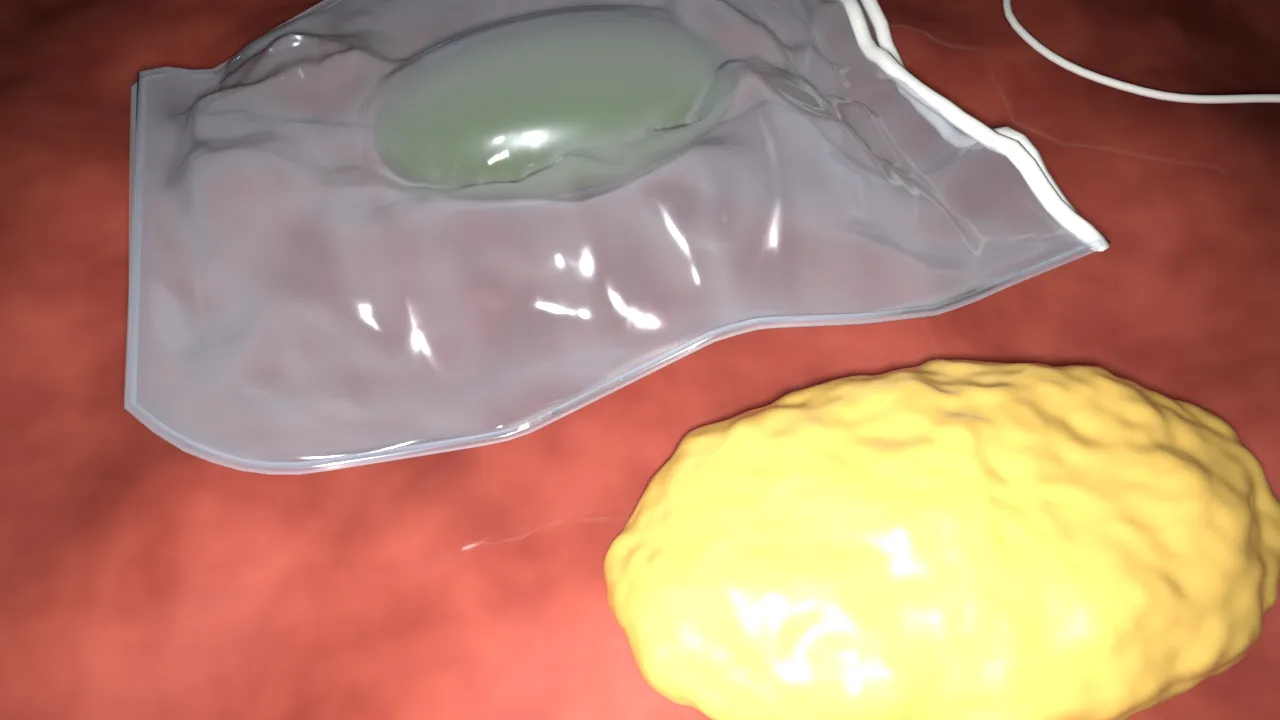
CVAT label: specimen_bag
At the end of the operation the gallbladder is put into a retrieval bag and removed through a port.
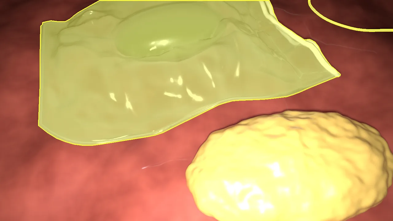
- Look for: a clear or milky plastic film crumpled into sharp creases, with thin streaks of glare along them. The edge has a welded seam, and the mouth usually has a hem with a drawstring or a stiffer rim.
- Do not confuse with: glare on wet tissue or smoke. The bag has straight creases and a clean edge; glare does not. Tissue seen through the film keeps its colour, only a little paler.
- The mask (proposed): the whole bag as one shape, including what you see through the film, plus the drawstring as far as you can follow it. This rule is still waiting for the Clinical Lead, so follow your CVAT task if it says otherwise.
specimen_bag colour.Telling them apart
| What you see | Most likely | Check |
|---|---|---|
| Two straight jaws, often with a window | grasper |
Jaws all metal, opening symmetrically |
| Slender curved jaws with coloured insulation | bipolar |
Bare metal only at the very tips |
| A thin wire bent through a right angle | hook |
Tapered insulated shaft behind it |
| Two thin blades crossing at a pivot | scissors |
Blades slide past each other, no window |
| A wide shaft, broad jaws, a clip between them | clipper |
Shaft visibly wider than the others |
| A blunt tube with side holes | irrigator |
No jaws, no bend |
| Crumpled clear film with streaks of glare | specimen_bag |
Straight creases, a seam at the edge, a drawstring or rim at the mouth |
| Only a shaft, the tip out of view | Unknown | Neighbouring frames, or tag disputed |
Open questions for the Clinical Lead
- The project’s annotation guideline v1.0 mentions separate labels for shaft, joint and jaws, while the CVAT label set has one class per instrument. Until this is settled, follow the label set of your task.
- Whether the specimen bag mask includes what is seen through the film, and the drawstring.
- How to draw edges blurred by motion or veiled by smoke.
- Whether clips placed on tissue get a class of their own.
About the pictures and models
The renders and models come from scripts/blender/instruments.py in the website repository and are published under CC BY 4.0, like the site’s other illustrations. The tissue bed is noise, not anatomy. The masks are rendered with one sample per pixel, so every pixel is wholly in or out of the instrument. Download a model: grasper, bipolar, hook, scissors, clipper, irrigator, specimen bag (glTF binary).
Back to annotation in CVAT.